


A spore trap collects airborne particles onto a surface for microscopic examination, but a slide count alone does not establish air concentration. Agricultural users need the actual sampled air volume, the fraction of the deposit examined, and a defensible identification method. Camera images cannot establish whether every captured particle is a viable crop pathogen.
How does a spore trap produce a slide for microscopy?
Follow the spore trap sample rather than the dashboard. In an active impaction arrangement, a pump draws air through an inlet and particles strike a prepared collection surface. A microscope then reveals deposited objects. Other collection arrangements require their own interpretation; a passive settling slide does not provide the same known air volume as an actively pumped sample.
Define the intended observation before choosing hardware. A crop investigation might need a count of a recognizable spore group over a stated collection period. That is different from identifying a pathogen to species, demonstrating viability, or estimating infection probability. Ask which of those tasks the collection method and microscopy procedure can actually support for the target crop.
Keep the collection surface, inlet geometry and preparation procedure together as one method. Adhesive thickness, contamination and particle loading can change whether material is captured and visible. A clean image does not prove a representative airborne sample. Conversely, a valid collection can become unusable if the slide is damaged or its identity is lost during transport.
Request a demonstration using the proposed collection surface and the actual target material. Record the slide identifier, sampling start and stop, measured flow, collection location and examiner. Retain an untreated blank when the agreed procedure requires one. A blank can reveal handling contamination, but subtracting its count mechanically from every sample can create another unsupported result.
Calculate sampled air volume before reporting spores per cubic metre
Use this illustrative calculation based on assumed site values, not a measured field record. Assume verified constant flow of 12 L/min and a complete, uninterrupted collection. Air volume in cubic metres equals flow in litres per minute multiplied by elapsed minutes and divided by 1,000. These are calculation inputs, not specifications for a NiuBoL sampler.
| Collection duration | Assumed flow | Sampled volume | Condition |
|---|---|---|---|
| 5 min | 12 L/min | 0.060 m³ | Constant verified flow |
| 10 min | 12 L/min | 0.120 m³ | Constant verified flow |
| 15 min | 12 L/min | 0.180 m³ | Constant verified flow |
| 20 min | 12 L/min | 0.240 m³ | Constant verified flow |
| 25 min | 12 L/min | 0.300 m³ | Constant verified flow |
| 30 min | 12 L/min | 0.360 m³ | Constant verified flow |
A 10 min collection at 12 L/min samples 0.120 m³, because 12 × 10 / 1,000 = 0.120. If the complete deposit contains 24 counted target objects, dividing 24 by 0.120 gives 200 spores/m³. Report that as an estimate from the collected sample, not an efficiency-corrected concentration or a measured infection risk.
Doubling collection to 20 min gives 0.240 m³; an unchanged concentration would yield an expected count of 48 under unchanged collection conditions. That expectation is arithmetic, not a promise about the next sample. Biological variation and collection losses remain. Longer sampling may also crowd the surface until additional objects are harder, rather than easier, to distinguish.
A pump outage of 2 min during a scheduled 10 min period leaves 8 min of active collection. At the assumed flow, volume becomes 0.096 m³ rather than 0.120 m³. Dividing the same 24 objects by 0.096 gives 250 spores/m³. Confirm the interruption record before calculating; do not infer active duration from the upload timestamp.
If flow changes, add the separately measured segment volumes. For example, 6 min at 12 L/min plus 4 min at 9 L/min gives 0.108 m³. The corresponding estimate from 24 objects is approximately 222.2 spores/m³. A missing flow history cannot be repaired by choosing a convenient average after seeing the count.
What changes when only part of the slide is counted?
A spore trap microscopy worksheet must say whether the examiner counted the entire deposit, predetermined traverses, or selected microscope fields. Fields chosen because they look busy are not a defensible area sample. Write the counting path before examination, include clear edge rules, and record how the observed area relates to the deposited trace.
For another illustrative calculation based on assumed site values, not a measured field record, assume a uniformly distributed deposit. Examining 25% of the area and observing 18 objects gives an expanded estimate of 18 / 0.25 = 72 objects. With a sampled volume of 0.180 m³, the calculated result is 400 spores/m³. The factor of four is an area expansion, not a correction for particles that bypassed the collection surface.
Reducing the examined fraction to 10% makes each counted object represent ten deposited objects under that assumption. At 0.180 m³, one additional observed object changes the expanded result by approximately 55.6 spores/m³. Such sensitivity matters when sparse observations are reported with decimal places. More digits do not resolve uncertainty about distribution across the slide.
Do not expand an arbitrary crop of an image as though it were a validated fraction of the deposition area. Trace edges, particle size and impaction patterns can produce nonuniform deposits. Compare counts from different parts of the trace during method evaluation. If the pattern invalidates the area assumption, examine more material or report the observed count without an unjustified concentration.
Camera resolution is not microscope identification capability
Request a calibrated scale at the specimen plane. Magnification printed on a lens does not state how many image pixels represent the smallest feature needed for identification. Focus, optical resolution, illumination and preparation also matter. A distant crop camera and a specimen microscope are different instruments, even when both supply a remotely accessible photograph.
The following example is an illustrative calculation based on assumed site values, not a measured field record. It uses a 2,400-pixel image width to show sampling geometry only. No row establishes optical resolving power, taxonomic identification accuracy or a specification for a commercial instrument. Match the proposed field width to a calibration slide rather than relying on the camera's total pixel count.
| Assumed specimen field width | Image width | Sampling pitch | Pixels across an assumed 6 µm object |
|---|---|---|---|
| 0.6 mm | 2,400 pixels | 0.25 µm/pixel | 24 pixels |
| 0.9 mm | 2,400 pixels | 0.375 µm/pixel | 16 pixels |
| 1.2 mm | 2,400 pixels | 0.50 µm/pixel | 12 pixels |
| 1.8 mm | 2,400 pixels | 0.75 µm/pixel | 8 pixels |
| 2.4 mm | 2,400 pixels | 1.00 µm/pixel | 6 pixels |
A 1.2 mm field across 2,400 pixels gives 0.50 µm/pixel after converting millimetres to micrometres. An assumed 6 µm object therefore spans 12 pixels. Widening the field to 2.4 mm reduces that span to 6 pixels. Confirm that diagnostic structures are visible; object width alone cannot demonstrate that similar-looking taxa can be separated.
A spore trap camera also has a depth limitation. Material outside the focus plane may be present but unreadable, while overlapping particles can obscure diagnostic features. Repeated photographs of an unchanged deposit do not create independent air samples. Preserve the original slide where practical and permit an examiner to return to ambiguous objects instead of forcing every image into a confident species label.
Place the sampler where the crop question can be answered
Record the inlet position relative to the crop canopy and nearby obstructions. A point beside a dusty access road may collect material that is not representative of the target block. A location above the canopy answers a different exposure question from a sheltered position among leaves. Keep the position fixed for a trend, or mark a relocation as a method change.
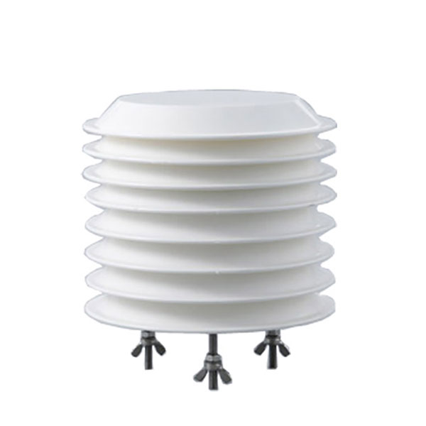
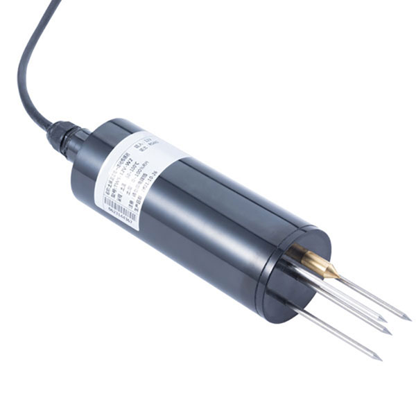
Use separate environmental observations to interpret collection conditions. The NiuBoL leaf wetness and temperature sensor provides crop-surface context; it does not count airborne spores. The combined air temperature and relative humidity sensor describes air conditions, not fungal identity. Align those records with the collection interval rather than attaching the latest available weather value.
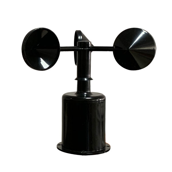
A cup wind speed sensor can add local airflow context, although wind speed alone cannot identify the origin of a deposited particle. Specify the exposure of each instrument separately. For NiuBoL agricultural monitoring projects, require the spore sampling method as a distinct deliverable rather than assuming an insect imaging cabinet supplies microscopic pathogen observations.
Reject results when volume, visibility or identity is missing
Do not use a spore trap concentration when the sampled volume cannot be reconstructed. An inlet restriction, an unrecorded pump stop or an incorrectly fitted collection surface can invalidate the denominator. A connected cabinet only establishes communication. Inspect collection and flow records before interpreting a flat trace as an absence of airborne material.
An unreadable or overloaded slide is not a zero. Record a visibility limitation, retain the image, and repeat collection under an agreed adjustment if the method permits it. Shortening a collection changes its temporal coverage, so preserve the revised duration. Never combine a shortened sample count with the longer scheduled volume to make a trend look continuous.
Microscopic appearance does not establish viability, and some organisms cannot be distinguished reliably by morphology alone. Use an appropriate specialist identification or separate analytical method when the decision requires more. A detected spore group does not prove active disease in the crop. Inspect susceptible tissue and relevant environmental conditions before deciding whether a treatment investigation is warranted.
Spore trap questions about slides, volume and images
Can a passive slide report spores per cubic metre?
Not from exposure duration alone. A passive slide records deposited material over an area and time, without a directly measured pumped air volume. Report the quantity supported by its collection procedure. Do not insert an assumed active-sampler flow into a passive-slide calculation or compare its count directly with a pumped concentration.
How do I calculate concentration from a whole-slide count?
Divide the counted target objects by the verified sampled volume in cubic metres. In the assumed example, 24 objects divided by 0.120 m³ gives 200 spores/m³. State whether the whole deposit was examined and whether collection efficiency is uncorrected. Keep the original count and volume so another examiner can reproduce the calculation.
Can I multiply a quarter-slide count by four?
Only when the examined quarter is representative under the adopted counting method. Eighteen objects in an assumed representative 25% area expand to 72 deposited objects. A dense patch selected for convenience does not meet that assumption. Predetermine the traverses, inspect deposit uniformity, and retain the examined fraction in the result record.
Will a larger camera identify smaller spores?
Not necessarily. More pixels help only when the optics, specimen scale and focus preserve useful detail. An assumed 6 µm object spanning 12 pixels still may lack visible diagnostic features. Request calibrated specimen images of the relevant targets and an explanation of unresolved groups, rather than accepting total camera resolution as identification evidence.
Does a positive slide mean the crop needs spraying?
No. Captured airborne material does not establish viable infection, tissue damage or an economic treatment threshold. Confirm the organism as far as the method allows, examine the crop, and consider susceptibility and weather. Use locally appropriate agronomic guidance for action. Preserve a distinction between a sampling observation and a disease diagnosis.
What information should accompany an unreadable sample?
Keep its identifier, collection interval, flow record, inlet position, slide preparation and image. Explain whether crowding, debris, poor focus or damage prevented examination. Report the result as limited or invalid according to the agreed procedure, not zero. A replacement sample should receive a new identity and retain its own volume and duration.
A count becomes usable when its denominator is visible
A spore trap result needs a traceable slide, measured air volume and a stated examined fraction. Under the assumed whole-deposit calculation, 24 objects in 0.120 m³ give 200 spores/m³, without correcting collection losses or proving viability. Before ordering, ask for a complete sample record and demonstrate how ambiguous images and interrupted collections are reported.
Send your crop, target spore group and proposed collection interval so the sampling method, microscopy work and separate environmental sensors can be discussed as distinct requirements.